Saturday, May 5, 2007

Secretin Testing

Secretin
From Wikipedia, the free encyclopedia

Secretin
Identifiers
Symbol
SCT
Entrez
6343
HUGO
10607
OMIM
182099
RefSeq
NM_021920
UniProt
P09683

Other data
Locus
Chr. 11 p15.5
Secretin is a peptide hormone produced in the S cells of the duodenum in the crypts of Lieberkühn.[1] Its primary effect is to regulate the pH of the duodenal contents via the control of gastric acid secretion and buffering with bicarbonate. It was the first hormone to be identified (see Discovery). In humans, the secretin peptide is encoded by the SCT gene.[2][3]

Discovery
In 1902, William Bayliss and Ernest Starling were studying how the nervous system controls the process of digestion.[4] It was known that the pancreas secreted digestive juices in response to the passage of food into the duodenum. They discovered (by cutting all the nerves to the pancreas in their experimental animals) that this process was not, in fact, governed by the nervous system. They determined that a substance secreted by the intestinal lining stimulates the pancreas after being transported via the bloodstream. They named this intestinal secretion secretin. Secretin was the first such "chemical messenger" identified. This type of substance is now called a hormone, a term coined by Bayliss in 1905.

Structure
Secretin is a linear peptide hormone, which is composed of 27 amino acids and has a molecular weight of 3055. A helix is formed in the amino acids between positions 5 and 13. The amino acids sequences of secretin have some similarities to that of glucagon, vasoactive intestinal peptide (VIP), and gastric inhibitory peptide (GIP). Fourteen of 27 amino acids of secretin reside in the same positions as in glucagon, 7 the same as in VIP, and 10 the same as in GIP.[5]
Secretin also has an amidated carboxyl-terminal amino acid which is valine.[6] The sequence of amino acids in secretin is: His-Ser-Asp-Gly-Thr-Phe-Thr-Ser-Glu-Leu-Ser-Arg-Leu-Arg-Asp-Ser-Ala-Arg-Leu-Gln-Arg-Leu-Leu-Gln-Gly-Leu-Val(NH2).[6]

Physiology


  • Production
    Secretin is synthesized in cytoplasmic secretory granules of S-cells which are found mainly in mucosa of duodenum, and smaller numbers in jejunum of small intestine.[7]
  • Stimulus
    Secretin is released into circulation and/or intestinal lumen in response to low duodenal pH that ranges between 4 and 4.5 depending on species.[8]
    It is the active form of prosecretin.This acidity is due to chyme, which contains hydrochloric acid, entering from the stomach via the pyloric sphincter.Secretin targets the pancreas, which cause the organ to secrete a bicarbonate-rich fluid that flows into the intestine. Bicarbonate ion is a base which neutralizes the acid, thus establishing a pH favorable to the action of other digestive enzymes to the small intestine and preventing acid burns[9] Other factors are also involved in the release of secretin such as bile salts and fatty acids which result in additional bicarbonate being added to the small intestine.[10] Secretin release is inhibited by H2 receptor antagonists which reduce gastric acid secretion. As a result, the pH in the duodenum increases above 4.5, and secretin cannot be released.[11]
  • Function
    Secretin stimulates the secretion of bile from the liver. It also increases watery bicarbonate solution from pancreatic duct epithelium. Pancreatic acinar cells have secretin receptors in their plasma membrane. As secretin binds to these receptors, it stimulates adenylate cyclase activity and converts ATP to cyclic AMP.[12] Cyclic AMP acts as second messenger in intracellular signal transduction and leads to increase in release of watery carbonate.It is known to promote the normal growth and maintenance of the pancreas.
    Secretin increases water and bicarbonate secretion from duodenal Brunner's glands in order to buffer the incoming protons of the acidic chyme.[13] It also enhances the effects of cholecystokinin to induce the secretion of digestive enzymes and bile from pancreas and gallbladder, respectively.
    It counteracts blood glucose concentration spikes by triggering increased insulin release from pancreas, following oral glucose intake.<[14]
    It also reduces acid secretion from the stomach by inhibiting gastrin release from G cells.[citation needed] This helps neutralize the pH of the digestive products entering the duodenum from the stomach, as digestive enzymes from the pancreas (eg, pancreatic amylase and pancreatic lipase) function optimally at neutral pH.[citation needed]
    In addition, secretin simulates pepsin secretion which can help break down proteins in food digestion. It also stimulates release of glucagon, pancreatic polypeptide and somatostatin.[8]
Uses
Secretin has been widely used in medical field especially in pancreatic functioning test. Secretin is either injected[15] or given through the tube that is inserted through nose, stomach then duodenum.[16] This test can provide information whether there are any abnormalities in pancreas which can be gastrinoma, pancreatitis or pancreatic cancer.
Extensive research has been conducted on the use of secretin to treat Autism. A "gut-brain" theory of autism proposes a link between the gastrointestinal disorders observed in many children with autism and their brain dysfunctions.[17]

References

  1. ^ Häcki WH (September 1980). "Secretin". Clin Gastroenterol 9 (3): 609–32. PMID 7000396.
  2. ^ Kopin AS, Wheeler MB, Leiter AB (March 1990). "Secretin: structure of the precursor and tissue distribution of the mRNA". Proc. Natl. Acad. Sci. U.S.A. 87 (6): 2299–303. PMID 2315322. PMC: 53674. http://www.pnas.org/cgi/pmidlookup?view=long&pmid=2315322.
  3. ^ Whitmore TE, Holloway JL, Lofton-Day CE, Maurer MF, Chen L, Quinton TJ, Vincent JB, Scherer SW, Lok S (2000). "Human secretin (SCT): gene structure, chromosome location, and distribution of mRNA". Cytogenet. Cell Genet. 90 (1-2): 47–52. PMID 11060443. http://content.karger.com/produktedb/produkte.asp?typ=fulltext&file=ccg90047.
  4. ^ Bayliss W, Starling EH (1902). "The mechanism of pancreatic secretion". J. Physiol. (London) 28: 325–353.
  5. ^ Williams, Robert L. (1981). Textbook of Endocrinology. Philadelphia: Saunders. pp. 697. ISBN 0-7216-9398-9.
  6. ^ a b DeGroot, Leslie Jacob (1989). J. E. McGuigan. ed. Endocrinology. Philadelphia: Saunders. pp. 2748. ISBN 0-7216-2888-5.
  7. ^ Polak JM, Coulling I, Bloom S, Pearse AG (1971). "Immunofluorescent localization of secretin and enteroglucagon in human intestinal mucosa". Scandinavian Journal of Gastroenterology 6 (8): 739–44. PMID 4945081.
  8. ^ a b Frohman, Lawrence A.; Felig, Philip (2001). "Gastrointestinal Hormones and Carcinoid Syndrome". in P. K. Ghosh and T. M. O’Dorisio. Endocrinology & metabolism. New York: McGraw-Hill, Medical Pub. Div. pp. 1326. ISBN 0-07-022001-8.
  9. ^ http://www.vivo.colostate.edu/hbooks/pathphys/endocrine/gi/secretin.html
  10. ^ Osnes M, Hanssen LE, Flaten O, Myren J (March 1978). "Exocrine pancreatic secretion and immunoreactive secretin (IRS) release after intraduodenal instillation of bile in man". Gut 19 (3): 180–4. PMID 631638. PMC: 1411891. http://gut.bmj.com/cgi/pmidlookup?view=long&pmid=631638.
  11. ^ Rominger JM, Chey WY, Chang TM (July 1981). "Plasma secretin concentrations and gastric pH in healthy subjects and patients with digestive diseases". Digestive diseases and sciences 26 (7): 591–7. PMID 7249893.
  12. ^ Gardner JD (1978). "Receptors and gastrointestinal hormones". in Sleisenger MH, Fordtran JS. Gastrointestinal Disease (2nd edition ed.). Philadelphia: WB Saunders Company.
  13. ^ Hall, John E.; Guyton, Arthur C. (2006). Textbook of medical physiology. St. Louis, Mo: Elsevier Saunders. pp. 800–801. ISBN 0-7216-0240-1.
  14. ^ Kraegen EW, Chisholm DJ, Young JD, Lazarus L (March 1970). "The gastrointestinal stimulus to insulin release. II. A dual action of secretin". J. Clin. Invest. 49 (3): 524–9. doi:10.1172/JCI106262. PMID 5415678.
  15. ^ "Human Secretin". Patient Information Sheets. United States Food and Drug Administration. 2004-07-13. http://www.fda.gov/cder/consumerinfo/druginfo/Human_Secretin.HTM. Retrieved on 2008-11-01.
  16. ^ "Secretin stimulation test". MedlinePlus Medical Encyclopedia. United States National Library of Medicine. http://www.nlm.nih.gov/medlineplus/ency/article/003892.htm#Definition. Retrieved on 2008-11-01.
  17. ^ "The Use of Secretin to Treat Autism". NIH News Alert. United States National Institutes of Health. 1998-10-16. http://www.nichd.nih.gov/news/releases/secretin.cfm. Retrieved on 2008-11-30.
See also
Secretin receptor

External links
Overview at colostate.edu
MeSH Secretin
Physiology at MCG 6/6ch2/s6ch2_17
[show]
vdeEndocrine system: hormones (Peptide hormones · Steroid hormones)
Endocrine glands
Hypothalamic-pituitary
Hypothalamus
GnRH · TRH · Dopamine · CRH · GHRH/Somatostatin
Posterior pituitary
Vasopressin · Oxytocin
Anterior pituitary
α (FSH, LH, TSH) · Prolactin · POMC (ACTH, MSH, Endorphins, Lipotropin) · GH
Adrenal axis
Adrenal cortex: aldosterone · cortisol · DHEAAdrenal medulla: epinephrine · norepinephrine
Thyroid axis
Thyroid: thyroid hormone (T3 and T4) · calcitoninParathyroid: PTH
Gonadal axis
Testis: testosterone · AMH · inhibin
Ovary: estradiol · progesterone · inhibin/activin · relaxin (pregnancy)Placenta: hCG · HPL · estrogen · progesterone
Other end. glands
Pancreas: glucagon · insulin · somatostatin
Pineal gland: melatoninThymus: Thymosin · Thymopoietin · Thymulin
Non-end. glands
digestive system: Stomach: gastrin · ghrelin · Duodenum: CCK · GIP · secretin · motilin · VIP · Ileum: enteroglucagon · Liver/other: Insulin-like growth factor (IGF-1, IGF-2)
Adipose tissue: leptin · adiponectin · resistin
Skeleton: Osteocalcin
Kidney: JGA (renin) · peritubular cells (EPO) · calcitriol · prostaglandinHeart: Natriuretic peptide (ANP, BNP)
Target-derived
NGF · BDNF · NT-3
[show]
vdeDigestive system, physiology: gastrointestinal physiology
Enteric nervous system
Meissner's plexus · Auerbach's plexus
Exocrine
Chief cells (Pepsinogen) · Parietal cells (Gastric acid, Intrinsic factor) · Goblet cells (Mucus)
Endocrine/paracrine
G cells (gastrin) · D cells (somatostatin) · ECL cells (Histamine)
enterogastrone: I cells (CCK) · K cells (GIP) · S cells (secretin)Enteroendocrine cells · Enterochromaffin cell · APUD cell
Border
Brunner's glands · Paneth cells · Enterocytes
Fluids
tract: Saliva · Gastric juice · Intestinal juiceaccessory: Bile · Pancreatic juice
Processes
upper GI: Swallowing · Vomiting
lower GI: Segmentation contractions · Migrating motor complex · Borborygmus · Defecation
either/both: Peristalsis (Interstitial cell of Cajal) · Gastrocolic reflexaccessory: Enterohepatic circulation
[show]
vdePeptides: neuropeptides
Hypothalamic
Somatostatin - CRH - GnRH - GHRH - Orexins - TRH - POMC (ACTH, MSH, Lipotropin)
Gastrointestinal hormones
Cholecystokinin - Gastric inhibitory polypeptide - Gastrin - Motilin - Secretin - Vasoactive intestinal peptide
Other hormones
Vasopressin - Calcitonin -
Other
Angiotensin - Bombesin/Neuromedin B - Calcitonin gene-related peptide - Carnosine - Delta sleep-inducing peptide - FMRFamide - Galanin - Gastrin releasing peptide - Kinins (Bradykinin, Tachykinins ) - Neuromedin (B, N, U) - Neuropeptide Y - Neurophysins - Neurotensin - Opioid peptide - Pancreatic polypeptide - Pituitary adenylate cyclase activating peptide
[show]
vdeHormones: gastrointestinal hormones
CCK - EGF - GIP - Gastrin releasing peptide - Gastrins - Proglucagon - Motilin - Peptide YY -Prokineticin - Secretin - VIP

Retrieved from "http://en.wikipedia.org/wiki/Secretin"
Categories: Genes on chromosome 11 Peptide hormones Intestinal hormones Digestive system

To view information on another disease, click on Pancreatitis SOD Library.!
Pancreatitis SOD Library

Barium X-rays, Ultrasound (Sonogram), Cat Scans, Nuclear Scans

Special Imaging Studies
...

Your doctor may have recently requested that you have an examination of your digestive tract at your hospital outpatient radiology (x-ray) department. If you are scheduled to have such an exam, it is important that you understand why the test is necessary and what steps are needed for proper preparation. This information reviews the most common studies performed. If you have any additional questions, please ask.
...
Why Do I Need This Test?
Tests such as these are done to provide your doctor with additional information about the status of your digestive system. This helps your doctor decide what, if any, problems may be present, and the best remedy in your particular case.
  1. Often, x-rays are requested to evaluate an organ that cannot be seen by other means. For instance, a diseased gallbladder may not show up in blood tests, nor can a standard "scope" test see this organ. But, specialized x-rays and other studies can visualize the gallbladder and determine if stones or disease are present.
  2. Sometimes x-rays are used to visualize part of the intestine that cannot be reached with a scope exam. A common example would be a barium x-ray of the portion of the small intestine that lies beyond the reach of a upper or lower scope test.
  3. Occasionally, a barium x-ray of the colon is ordered when severe diverticulosis or other problems prevent completion of a full colonoscopy "scope" examination.
..
If you need to have x-rays or other special studies performed, your doctor will explain what tests are necessary and why.
..
Here is a summary of the four basic exams:
  1. Barium X-rays You may not realize that when ordinary x-rays are taken, only your bones show up. X-ray beams pass right through the soft tissues of your body, such as the intestines, liver, kidneys, etc., making them almost invisible. Barium is an inert, harmless mineral that prevents the passage of x-rays - just as your hand blocks sunlight. During an Upper Gastrointestinal Series, or Upper GI Series, you are given a flavored "milkshake" of liquid barium to drink. The barium fills your stomach making it visible on the x-ray film. The standard Upper GI evaluates the esophagus, stomach, and first portion of your small intestine - called the duodenum. This takes about 30 minutes. If your doctor requests that the remaining twenty feet of small intestine also be evaluated, the test is termed a Small Bowel Series and takes an additional two to three hours to complete. Both examinations require fasting after midnight, but no laxative preparation is necessary, nor is a driver needed. A similar examination of the large intestine, or colon, is called a Lower Gastrointestinal Series, or Lower GI. Of course, in this instance, the barium is not swallowed, but is given rectally as an enema - thus the more common name, Barium Enema. This examination takes less than an hour and requires fasting as well as an unpleasant laxative and dietary preparation the day or two before. Prep instructions vary and will be provided by our staff. Be sure to follow them exactly so the test need not be repeated.
  2. Ultrasound Another test often performed in the x-ray department does not use x-rays at all - an Abdominal Ultrasound. Also known as a Sonogram, this test utilizes specialized sound waves that bounce off your internal organs to obtain images - much like the way a submarine uses sonar to locate the enemy. The pitch, or frequency, of these sound waves is far above the range of human hearing, hence the name Ultrasound. An Ultrasound is also the examination that is given to pregnant women to check the size of their baby before delivery. This same exam can also be used to visualize certain digestive organs, such as the gallbladder, liver and pancreas as well as the main abdominal blood vessel, the aorta. During an ultrasound, you will be positioned on an examination table and a gel will be applied to your abdomen. A small probe, called a transducer, will be passed over the surface of your abdomen. The test is quick, painless, and requires very little preparation. Fasting after midnight is usually required. A driver is not necessary. If your doctor also requests an evaluation of the lower abdomen, or pelvis, a full urinary bladder is also required.
  3. CAT Scans A CAT Scan, also known as CT Scan, uses a complex computerized x-ray scanner that takes multiple views of your abdominal organs as you lie on a flat table. A high speed computer analyses the information to create cross sectional images of parts of your body. The radiologist (x-ray doctor) can then view these images in sequence, like slices in a loaf of bread, and create a virtual three dimensional view of your abdominal organs. CT is best suited for viewing the more solid digestive organs like liver, pancreas and kidney, and less suited for viewing the hollow air field intestinal tract. Preparation is simple, however there are a few concerns. First, since dehydration can increase the risk of side effects, it is important that you drink plenty of fluids up until four hours before the examination. During the last four hours, you should avoid all food and drink. Since the contrast material can worsen previously existing kidney disease, the radiologist usually requests proof of normal kidney function. This requires a blood test. To make the examination more accurate, you will be asked to drink an oral contrast solution before the scan. Additional contrast material may also be given by vein to enhance the images. Since this solution contains iodine, be sure to tell the doctor if you have ever had an allergic reaction to iodine, IVP or catheterization dye, or shellfish. Depending on the nature of your previous reaction, the solution will have to be modified or simply not used. The CT scan itself is simple and painless, and usually takes less than an hour to complete. Serious side effects are rare, but a temporary feeling of warmth and mild nausea are common after the contrast injection. After the examination, you will be able to resume your normal activities and diet. You should drink plenty of fluids for 24 hours after the examination to help flush out your system.
  4. Nuclear Scans In some instances, your doctor may request that you have a nuclear scan. Although the name sounds a little unsettling, a nuclear scan involves only a small "tracer" dose of radioactive material, and is not dangerous. Once this tracer is injected into your system, it can be followed through your digestive organs as you lie directly underneath a large, flat Geiger counter. A nuclear scan is most often used to assess liver and gallbladder function. Other uses include measurement of stomach emptying and localization of intestinal bleeding. Nuclear scans require very little preparation.

..

Where Is The Test Performed?
X-ray, Ultrasound, CAT scan and Nuclear Scan exams require specialized equipment and highly trained technicians. They are usually performed in a hospital our specialized outpatient facility.
..
Who Performs The Exam?
In most cases, the examination will be performed by a specially trained technician who is expert in using the equipment. The radiologist may or may not be present depending on the circumstances. However, the radiologist will review the final films and proceed with an official evaluation.
..
How Do I Schedule My Test?
Your doctor's office can assist you in obtaining all the necessary information to schedule your examination with your local outpatient radiology department. Any special preparation instructions will be provided.
..
Preparing for Your
Test Careful preparation is an important part of any examination. Preparation instructions vary from test to test and will be provided by our office staff. In all instances, you should wear easily-removable and comfortable clothing. Leave girdles, valuables, high heels, and jewelry at home. Be sure that you fully understand what preparation is required of you so that a safe and accurate examination can be performed. If you do not, the examination may have to be repeated at a later date. If you have any questions, please ask.
..
After Your Exam
After your examination is completed, you will be able to immediately resume normal activities and diet. You should drink plenty of extra fluids for the first 24 hours to help flush your system.
..
A word to the wise: If barium was used for your test, you might also wish to take a mild laxative such as an ounce of Milk of Magnesia after the x-ray. This helps eliminate the barium from your system. If this is not done, the barium can harden, much like plaster of Paris, making elimination quite difficult.
..
What About Side Effects?
There are no known side effects to health from ultrasound. Nuclear scans, CAT scans, and barium x-rays do expose your body to small amounts of radiation. However, the dose is quite low and felt to be generally safe. The exceptions would be pregnancy and breast feeding. Any woman who is pregnant, or thinks she might be, or is breastfeeding, should let her doctor know before scheduling the examination. Also, if you believe that you are allergic to iodine dyes and are scheduled for a CT scan, notify your doctor. How Do I Get The Results? Once the test is completed, the films are developed and analyzed by the radiologist who then sends us a written report. Your doctor will contact you after reviewing the results in the context of your case. This may take about a week, but most reports arrive sooner. In Summary In addition to "scope" tests, imaging techniques such as barium x-rays, Ultrasound studies, CAT and Nuclear Scans are often requested to obtain information about the status of your digestive system. These tests are generally safe and well tolerated, but require proper preparation. You have an important role in making your examination a success. If you have any questions, again please ask.
..


To view information on another disease, click on Pancreatitis SOD Library.!

Pancreatitis SOD Library


..


Camera in a Pill


Wireless Capsule Endoscopy - "The Camera in a Pill"
Searching For the Leak.... What if you were a plumber and couldn't find the leak? That is the situation that sometimes frustrates doctors asked to evaluate a patient with signs of iron deficiency anemia due to blood loss. There are many types of anemia. Iron deficiency anemia is a common type and most often occurs in pregnant women or menstruating women due to the monthly loss of blood. But when significant iron deficiency anemia occurs in women after the age of menopause or in men, it is often a sign of the loss of blood from somewhere else - usually the digestive tract. This blood loss may be very gradual and not visible to the patient. But over time, the continued loss of blood can deplete the body of iron and anemia develops.
If a doctor suspects that anemia is due to intestinal bleeding, special tests are usually ordered to help the doctor "find the leak." This is a bigger task than most people realize since the average adult digestive tract is approximately 30 feet in length. The top 4 feet includes the esophagus (foodpipe) and stomach and first portion of the small intestine, called the duodenum. The bottom 6 feet makes up the colon and rectum. In between, lies the rest of the 20 feet of small intestine where the process of digestion actually occurs.
..
Scope Tests
In this situation, one of the first diagnostic studies ordered are special "scope" tests of the digestive tract. Gastroscopy is used to check the first 4 feet of the upper digestive tract and colonoscopy to evaluate the colon and rectum. As you can see, most of the 20 feet of small intestine lies beyond the reach of these two studies. Fortunately, most bleeding problems seem to occur in the area than can be "scoped" and the source of bleeding is usually found and treated. Common problems would include Hiatal hernia, gastritis, ulcers, polyps, and, sometimes, stomach or colon cancer.
..
The Small Intestine - Uncharted TerritoryWhat if a patient had severe iron deficiency anemia and scope tests of the stomach and colon are normal? It is not uncommon for doctors to evaluate a patient with unexplained anemia and neither gastroscopy nor colonoscopy scope examinations reveal the diagnosis. By a process of elimination, it then becomes likely that the source of bleeding lies somewhere in-between - below the reach of the gastroscope and above the reach of the colonoscope - in the 20 feet of small intestine. How then is this area examined?
Well, not very well. Gastroscopy and colonoscopy cannot reach this far. Contrary to popular belief, special imaging studies like CT scan or MRI are not useful in this circumstance. X-rays of the small intestine can be performed after drinking a chalky solution of barium. Called a small bowel series, this test has been available for many years, but has a limited accuracy. X-rays are still only shadow pictures and do not view the object itself like a camera.
..
Why not use a longer scope?
Since scope tests were first invented, doctors have wanted to be able to visualize the entire gut - all 30 feet. But, a direct view of the small intestine has remained elusive. Attempts have been made to develop longer endoscopic instruments. This technique called push enteroscopy has had only limited success. The longer instruments are difficult to control and manipulate and are hard to maintain. The accuracy of push enteroscopy is still limited since even in the best of hands the entire small intestine is not visualized.
..
The Given Video Capsule - Hope For the Future
In 1981, an Israeli physician, Dr. Gavriel Iddan, began development of a video camera that would fit inside a pill. Technology was not ready and the idea was put on hold. It took 20 years for technology to catch up with Dr. Iddan. In 2001, the FDA approved the Given Diagnostic Imaging System. This may sound like science fiction, but this 11 x 26 mm capsule weighs only 4 gms (about 1/7th of an ounce) and contains a color video camera and wireless radiofrequency transmitter, 4 LED lights, and enough battery power to take 50,000 color images during an 8-hour journey through the digestive tract. About the size of a large vitamin, the capsule is made of specially sealed biocompatible material that is resistant to stomach acid and powerful digestive enzymes. Another name for this new technique is Wireless Capsule Endoscopy.
..
Patients report that the video capsule is easier to swallow than an aspirin. It seems that the most important factor in ease of swallowing is the lack of friction. The capsule is very smooth, enabling it to slip down the throat with just a sip of water. After the Given M2A capsule is swallowed, it moves through the digestive track naturally with the aid of the peristaltic activity of the intestinal muscles. The patient comfortably continues with regular activities throughout the examination without feeling sensations resulting from the capsule's passage. During the 8 hour exam, the images are continuously transmitted to special antenna pads placed on the body and captured on a recording device about the size of a portable Walkman which is worn about the patient's waist. After the exam, the patient returns to the doctor's office and the recording device is removed. The stored images are transferred to a computer PC workstation where they are transformed into a digital movie which the doctor can later examine on the computer monitor. Patients are not required to retrieve and return the video capsule to the physician. It is disposable and expelled normally and effortlessly with the next bowel movement.
..
This technique is very new, but preliminary reports have already demonstrated cases in which the Given videocapsule was able to make a diagnosis not seen by conventional studies. This remarkable device will never be 100% accurate. No medical procedure is. But current estimates suggest an accuracy of about 55%, an improvement over push enteroscopy. As of May 2002, the software is still evolving and there are concerns over insurance coverage, but this remarkable device shows great promise. Currently, it is not available through our center or in Western Pennsyvania, but hopefully, it will be in the future. For more information about this device and its availability, access the Given Imaging Ltd. Website at:

www.givenimaging.com

To view information on another disease, click on Pancreatitis SOD Library.!

Pancreatitis SOD Library


..


Hemoccult Tests

Hemoccult Card Test (for hidden blood in the stool)

Tina: How a visit to her gynecologist saved her life.... Tina F. is a 52 year old school psychologist who sees her gynecologist every year for a routine checkup. Like many women, she does not have a family doctor, but uses her gynecologist as her personal physician. This year her doctor suggested that a routine stool Hemoccult test be performed in addition to her annual blood tests and physical exam. As she left the office, the nurse gave her a Hemoccult card test kit and instructions. Tina collected the stool specimens as instructed and mailed the card back to her doctor's office for testing. She was surprised to receive a letter from her doctor the next week informing her that the Hemoccult test found traces of hidden blood in her stool.
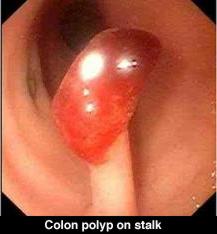
Tina was especially surprised since she never had any problems with bowel movements and never noticed any blood in her stool. She was referred to our office and a colonoscopy exam was performed to determine the cause of the bleeding. The colonoscopy exam went well and found the cause of Tina's bleeding - an inflamed colon polyp that was located in her right colon. It was slowly oozing small amounts of blood and was the source of the trace of hidden blood in the stool. In this series of photos, you can see how the polyp was removed during colonoscopy using a thin wire electrocautery snare. It was a simple outpatient procedure and she was back to work the next morning. Tina was pleased that her problem could be cured with a simple painless outpatient procedure and she was back to work the next morning. The polyp was sent for a biopsy.
We called Tina the next week and informed her that she was very lucky. The biopsy showed that her polyp was "highly dysplastic with carcinoma-in-situ bordering on adenocarcinoma" - about as close as you can get to cancer, without actually being malignant. Her gynecologist did her a great service by being thorough and discovering her rectal bleeding even before it was visually apparent. The discovery and removal of this type of colon polyp saved Tina from having to deal with colon cancer in her future and probably saved her life. She will be scheduled to return for a follow-up colon exam periodically in the future.
...
What is a Hemoccult test? The Hemoccult test, a product of SmithKline Diagnostics, Inc., is a simple screening test which detects blood in the stool (bowel movements).
It is a very sensitive test and can detect a small trace of blood even before it is visible to the naked eye. In medicine, the prefix "heme" refers to blood, as in "hematology". Blood which is hidden from view is called "occult" bleeding (nothing to do with witchcraft), hence the name Hemoccult for "hidden blood".

Other tests for occult blood are also on the market (Colocare, EZ Detect), but the most commonly used test is the Hemoccult.
..

Prevention - the purpose of the stool Hemoccult test is to help prevent colon cancer by searching for hidden (occult) blood in the stool before it has become so apparent to be visible. The American Cancer Society estimates that over 130,000 people developed colorectal cancer in 2000. Not only is this a common disease, it is quite sneaky. In fact, the majority of patients with colon cancer are incurable when they have their first symptom. Despite undergoing the rigors of heroic treatment such as surgery, radiation, and chemotherapy, most paitients succumb to their disease.
..Why do this test?In advanced stages, the overall cure rate is quite low. Over 56,000 Americans died last year from colon cancer. However, when colorectal cancer is detected in an early localized stage, up to 85% of colorectal cancers can be treated successfully. When cancer is detected after symptoms appear and it has spread to other parts of the body, fewer than 7% may be treated successfully. This means that earlier detection could save thousands of lives each year. In some cases, the Hemoccult test will show hidden blood in the stool in early curable stages of colon cancer. Even better, as in this case, the Hemoccult test may be positive even before cancer cells have developed. How is it done? It is quite simple. To complete the test, you place a thin smear of a small stool specimen, taken from your toilet bowl, onto the window inside the test slides included in your kit. You do this twice from each of three consecutive bowel movements and mail the completed card back to your doctor's office in the special envelope.
..
To maximize the accuracy, some special precautions are suggested:


  1. Do not collect samples during, or until three days after your menstrual period, or while you have bleeding hemorrhoids or blood in your urine.
  2. Remove any cleaning or deodorizing agents from the toilet and flush twice.
  3. For 3 days before the test, avoid Vitamin C tablets, iron tablets, red meat (rare, cooked and processed beef, lamb and liver), raw broccoli, cauliflower, horseradish, parsnips, radishes, turnips and melons.
  4. Protect slides from heat, light and chemicals. Keep cover flap of slides closed when not in use.
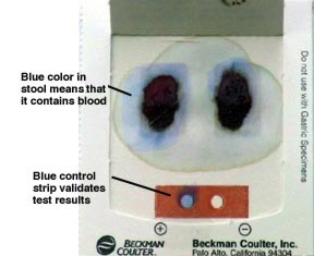
  5. To be sure the the card and developing solution are fresh and active, a quality control strip is included on the back of each card. Normally, the developing solution turns this control strip blue which means the card can be trusted.

What happens after the Hemoccult card is returned?

  1. After the stool collection is complete, the Hemoccult card is mailed back to the doctor's office or lab in a special protective envelope for testing.
  2. Testing the card is quite quick and simple. The medical technician opens the flaps on the back of the card and applies a few drops of a special chemical developing solution onto each of the three stool specimens.
  3. POSITIVE TEST The developer turns the stool specimen blue - this is an abnormal result and means that blood is present in that stool specimen.
  4. NEGATIVE TEST If the stool specimen does not turn blue, then no hidden blood is present. This is a normal result.


Who should do a Hemoccult test?

All adults over 40 should do this test annually even if there are no symptoms. This really should just be part of an annual checkup from your family doctor or gynecologist.

Who should NOT do a Hemoccult test?

Anyone with visible rectal bleeding. This test looks for hidden blood in the stool - before it is visible to the naked eye. If an individual has visible blood in the stool, there is no reason to do a Hemoccult test. You already know that bleeding is present and needs to be evaluated.

What if it is abnormal (POSITIVE)? Don't panic! A positive Hemoccult test (one that shows blood) does not diagnosis cancer. It simply means that there is blood in the stool that is not supposed to be there. There are many possible causes for blood in the stool which have nothing to do with colon cancer such as hemorrhoids, fissures, ulcers, and, as in the above example, colon polyps. The important point is that when a Hemoccult test is positive for occult blood, you must see your doctor for further testing to determine the cause. The safest approach is to assume that the cause is colon cancer until proven otherwise. Most adults who have an abnormal stool Hemoccult test will be referred to a gastroenterologist for further evaluation. As in Tina's case, this will usually include an examination of the colon, or colonoscopy. If the colon test is normal, the doctor will sometimes suggest that the upper digestive tract be evaluated in a similar fashion by also performing a gastroscopy test. Usually, both "scope" exams can be done on the same visit.

What if it is normal (NEGATIVE)? That sounds reassuring, but perhaps falsely so. The Hemoccult test is a test for blood, not cancer. The surface of a colon cancer is delicate and may bleed easily. In fact, this is the very basis of the test itself. But, you should know that most colon cancers and polyps bleed intermittently. They may lose a small amount of blood one day and not bleed again for several days. Some don't bleed at all - especially in early curable stages. So, a normal (NEGATIVE) stool Hemoccult test misses about 60% of colon polyps and 40% of colon cancers. Not very reassuring. This is really a screening test that has some significance only if abnormal (POSITIVE). The Hemoccult test is meant to be just a part of colon cancer prevention, but must be supplemented with more ac curate examinations of the colon, such as a screening sigmoidoscopy, a screening colonoscopy, or a barium enema.

How accurate is the Hemoccult test?

Not very. As mentioned above, the Hemoccult test is very sensitive for finding occult blood in the stool when present. It does so about 98% of the time. But since colon cancer and polyps may not bleed during the time of stool collection, a normal result may miss colon cancer and polyps up to 60% of the time. However, doing a Hemoccut test is better than doing nothing at all - which is what most adults do. The Hemoccult will still pick up about 40% of cases that would have been missed if no testing was done. However, the Hemoccult test should not be relied upon as the sole screening method.

Summary

The Hemoccult test is a simple, inexpensive, non-invasive stool test that can be collected in the privacy of your home. It is not a test for colon cancer, but detects hidden (occult) blood in the stool before it becomes visible. A normal test should not be reassuring since it misses over half of the colon cancers and polyps. It is not meant to be used alone, but must be part of a comprehensive colon cancer screening program which include other screening exams. A positive test should not cause panic, but must be further investigated to determine the source of bleeding. Only diagnostic testing from a physician can determine the cause of the bleeding. Used correctly, the stool Hemoccult test can be an important aid in detecting cancer in its early stages of development. If you have further questions, discuss them with your doctor.


To view information on another disease, click on Pancreatitis SOD Library.!

Pancreatitis SOD Library


..