Saturday, May 5, 2007

EUS

Endoscopic Ultrasound
..
What is Endoscopic Ultrasound (EUS)?
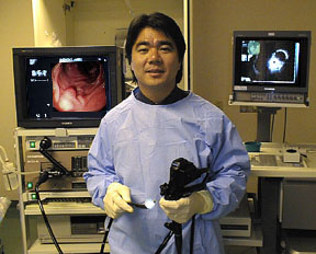
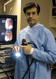
Before we discuss what Endoscopic Ultrasound is, it will be important to review what ultrasound means. Ultrasound is a technique that uses sound waves to create a picture of the inside of the body. In this way an ultrasound is similar to an x-ray, but no radiation is used. It is commonly used to see the fetus during pregnancy to determine the sex of the baby. Endoscopic ultrasound (EUS) is a technique that uses a special endoscope that has an ultrasound machine at the tip.


Using the EUS "scope", your doctor can place the ultrasound probe in the gastrointestinal (GI) tract, very close to the area of interest, so that the best possible picture can be obtained. EUS has been shown to be superior to routine x-rays such as abdominal ultrasound, barium studies, CAT scans, and even MRI scans for looking at the local spread of GI cancers, for evaluating lesions in the GI tract that are below the surface,
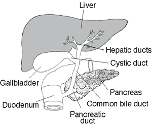
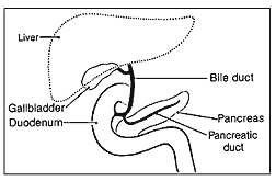
and for evaluating the pancreas and even the bile ducts that drain the liver. This procedure is very highly specialized and is performed by a gastroenterologist who has specialty training in this area.
..
How is EUS Performed?
To the patient, an EUS is very similar to an endoscopy or "scope" test. EUS can be performed in the upper GI tract or in the lower GI tract. Since you will receive sedation to make the exam painless, you must bring a companion with you to drive you home. For the Upper GI tract EUS exam, there is very little preparation except fasting after midnight the night before the test. You may drink clear liquids up to six hours prior to your exam, then nothing by mouth (not even water).Your usual medications may be taken with small sips of water. However, if you take blood thinners, aspirin or arthritis medicines such as Motrin, ibuprofen, etc.., you should let your doctor know, as these may need to be stopped before the test. In addition, if you are diabetic, you should also let your doctor know, so that adjustments in your doses can be made for the day of the test. Finally, if you have a history of a heart murmur, rheumatic fever as a child, infection of a heart valve, or an artificial heart valve, you should let your doctor know so that he can determine if antibiotics need to be ordered before the test. Just before the test, you will receive some medications in your IV that will make you sleepy and very relaxed. Usually, a regular "scope" is first performed in order to see the area of interest.
The doctor will carefully guide the scope into your mouth, through your food pipe, past the stomach, and into the first part of the small intestine, called the duodenum. After this scope is removed, the EUS scope is passed down in the same manner. The area of interest is then viewed with the ultrasound probe at the tip of the EUS scope. For the Lower GI tract EUS exam, you may also be asked to take 2 fleets enemas on the morning of the exam. Your doctor will then guide the EUS scope through the rectum to the area of interest. This test is generally very quick and is done without any medications, similar to a flexible sigmoidoscopy.
..
What about Biopsies or Sampling Tissue?
Occasionally, EUS reveals a lesion that needs to be sampled. Most of the time, this can be performed during the EUS examination. Your doctor may take a biopsy sample using a forcep or needle. This is generally performed while directly looking at the lesion through the "scope" or occasionally, this can be performed using the endoscopic ultrasound image as a guide.
..
What are the Risks of EUS?
The risks of EUS are similar to the risks associated with standard endoscopy. While every medical procedure involves some degree of risk, the frequency of complications during endoscopy is extremely low. The major risks are perforation, or creating a hole in the lining of the GI tract, and bleeding. If perforation should occur, it usually requires surgery to repair it. Bleeding may occur at the site of a biopsy or needle sample. Typically minor in nature, bleeding can generally be treated during the endoscopy by injecting medications or cauterizing the bleeding site, but rarely, surgery is required. Fortunately, perforation and bleeding are quite rare. Other minor risks of EUS include drug reactions and complications related to other diseases that you may have. Consequently, you should inform your doctor of all allergic tendencies and medical problems. Occasionally, the site of the sedative injection may become inflamed and tender. This usually is not serious and responds to warm compresses.
..
After the Test
After outpatient EUS, you will rest awhile in the recovery area. You may feel slightly bloated from the air that was placed in your GI tract, but this will gradually subside. You may have a sore throat which will also subside. Some people develop nausea due to the sedative medications given. Once you are more alert, your doctor will discuss the test's findings with you. If a biopsy was obtained, the doctor will contact you with the results when they become available. Since the effects of the sedative medication may take up to 24 hours to wear off completely, you should plan to go directly home. You will need to have a driver come with you as you will not be allowed to drive for the remainder of the day. Have a light meal and rest for several hours. After sedation has worn off, you may resume your regular diet, but you will not be able to return to work, drive, or operate any dangerous machinery for the remainder of the day. If you did not receive any sedation, you will be allowed to resume your normal activities at discharge.

To view information on another disease, click on Pancreatitis SOD Library.!

Pancreatitis SOD Library


..

Colonoscopy

Colonoscopy
..
Tremendous progress in the field of fiberoptics has made it relatively simple for your doctor to examine the entire large intestine, or colon, with a procedure called colonoscopy. More accurate than a barium enema x-ray and much simpler than exploratory surgery, colonoscopy is a safe and highly effective diagnostic technique. After careful assessment, your doctor has decided that a colonoscopy is necessary to better evaluate and treat your condition. Since your cooperation is essential for a successful examination, it is important that you understand exactly what is to be done and why. If you have any additional questions or concerns after reading this information, please ask your doctor.
..
What Is Colonoscopy?
Colonoscopy is a medical term that has two parts - colono which refers to the colon, or large intestine, and scopy which means "looking into." Therefore, colonoscopy is a test that enables your doctor to look inside your colon.
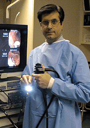
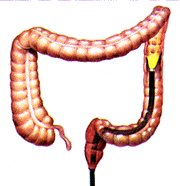
The instrument used to perform this test is the colonoscope: a long, flexible tube about the width of your index finger. Within the end of this tube is a miniaturized color-TV camera with a wide-angle lens. After passing this "scope" through the rectum and into your colon, your doctor then directly examines the lining of your lower digestive tract on a television monitor. In this manner, it is possible to evaluate intestinal inflammation, ulceration, bleeding, diverticulitis, colitis, colon polyps, tumors, etc. As part of your assessment, you may have undergone a barium enema examination of the colon, or "lower GI series." This technique, which utilizes x-rays, is helpful in identifying any areas of possible abnormality. X-rays are only shadows, however, and often do not fully demonstrate what is wrong. For example, the barium enema x-ray may miss nearly forty percent of colon polyps.
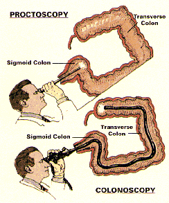
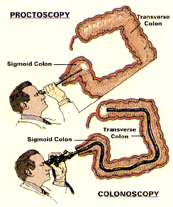
In the past, you also may have undergone a proctoscopic examination of the lower colon and rectum. Performed with a short, rigid, hollow tube, this limited procedure enabled the doctor to examine only the last ten inches of the large intestine. In most offices, the proctoscope has now been replaced by the flexible sigmoidoscope. This newer device enables your doctor to examine the last two feet of the colon while its flexibility affords the patient much greater comfort than does the rigid type of scope. The colonoscope offers a still greater advantage. With this longer flexible instrument, your doctor is usually able to directly examine the entire length of your colon - all five feet of its numerous twists and turns. In this manner, your doctor can be most certain about the condition of your colon.
..


How You Can Help
Your cooperation is essential for a successful examination. There are certain things you can do to help before the test begins:
  • Preparation A colonoscopy exam requires a thorough cleansing of the large intestine so that nothing impedes your doctor's view. The colon must be completely free of all solid waste. Since particles of stool can interfere with the examination, you will require a special dietary and laxative preparation on the day prior to the test. To be sure that you fully understand all aspects of the preparation, the GI Assistant or a member of the Nursing staff will review the preparation with you. It is critical that you follow the instructions as given. If the colon is not clean, the test - as well as the preparation - may have to be rescheduled. A few hints: It is best not to be constipated before the laxative preparation. This may lead to a temporary backup of the laxative solution, cramps, bloating, nausea, and vomiting. If you tend to be constipated, take one or two ounces of Milk of Magnesia the day before the laxative prep to "open up your pipes." This will make the actual preparation easier. If you develop mild distress during your prep, simply stop it for an hour or two and then resume it where you left off. If you develop severe abdominal cramps or persistent nausea and vomiting, stop the prep and call our office to reach our doctor on call. Of course, the laxative preparation will cause you to have many bowel movements. This may cause some rectal irritation and a small amount of bleeding. Many patients find that using Charmin Plus toilet tissue minimizes such rectal irritation. A nonprescription 1% hydrocortisone cream may also be soothing when applied to the rectal area after each bowel movement. Please avoid foods with many small seeds for a few days before the exam. They tend to linger in the colon and can clog our instruments. In addition, you must avoid all red-colored Jello products during your preparation as their red dye masks the lining of the colon.
  • Medications Medications containing iron or Metamucil-like fiber can impair your doctor's view of the colon. If possible, they should be temporarily discontinued several days before the test. If you are taking chronic aspirin or Persantine therapy, diabetic medication, or the blood-thinner Coumadin, you will require special preparation and adjustment of your medicines before the test. Please call our office for instructions. All other routine medications may continue to be taken with a small sip of water, even on the day of the exam.
  • What To Wear You will have to change into a patient gown before the examination. If this test is being done on an outpatient basis, you should wear, loose, comfortable, casual clothing that is easily removed and folded. Avoid girdles, pantyhose, or tight-fitting garments. Please leave your jewlery, valuables, and high heels at home. Transportation To minimize any discomfort, you will receive an injection before the colonoscopy. As this medication will make you drowsy for several hours, you cannot safely drive a car for the remainder of the day. Therefore, if this test is being done on an outpatient basis, a family member or friend must accompany you to the office in order to drive you home. If possible, you should choose someone with whom the doctor can freely discuss the results of your test. We request that your companion remain in the office during the test. You should arrive several minutes before your appointment and plan to stay approximately two hours. If the test is being done on an inpatient basis, a driver will not be needed. After the test, you will simply return to your hospital room to sleep off the remainder of the medication. You will be asked to remain in bed several hours to allow the sedation to wear off completely.
  • Your Permission If you have any questions or concerns about this test, do not hesitate to ask your doctor about them. To signify that you completely understand what this test involves, you will be asked to sign a written consent form, or "permit," before the test begins.

Looking Inside
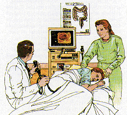
Prior to the test, you will be asked to remove your clothing and to slip into a patient gown. After signing the permission form, you will be positioned comfortably on your left side on the padded colonoscopy table. A small painless oximeter probe will be placed on your fingertip to monitor your pulse rate and breathing function during the exam. A thin nasal oxygen tube may be used to administer low flow oxygen during the exam. Then you will be given an intravenous injection of Demerol (pain killer) and Versed (tranquilizer); these medications will make you drowsy and relaxed, thus minimizing any discomfort during the examination. Please inform the doctor if you are allergic to Demerol or Versed, or if you chronically take narcotic painkillers such as Percodan, Percocet or Codeine, so that other medications can be substituted. Furthermore, rest assured that these medications will not prompt you to act or speak foolishly during the test.
..
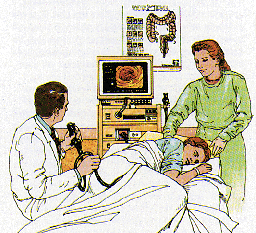
Your doctor will then examine and lubricate the rectal opening to allow simple passage of the colonoscope. This will not be painful, but is mildly uncomfortable for a few seconds. After the colonoscope is properly positioned, your doctor gently guides the scope through the colon, a process that takes approximately one-half hour. To improve visualization, your doctor gently infuses air into the colon. This may cause a sensation of abdominal fullness, but should not be painful. In fact, many patients actually fall asleep during the examination. As the test progresses, you may be asked to change your position now and then to facilitate the passage of the colonoscope through the colon.
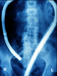
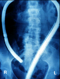
Here is an xray of the abdomen taken during a colonscopy which demonstrates how far the scope travels into the colon all the way to the right side of the colon.
..
Occasionally, passage of the colonoscope through the entire colon is impossible. While this may be caused by a blockage, it is most often caused by an extra "loop" of colon, adhesions from previous abdominal surgery, or severe diverticulosis. In such cases, a limited examination may be sufficient if the area of suspected abnormality has been visualized. If not, a barium enema x-ray (lower GI) may also be necessary.
..
Taking Specimens
If colonoscopy reveals any unusual condition such as inflammation, an ulcer, a tumor, or a polyp, your doctor may photograph it. This photo provides a permanent record for your medical chart and allows your other doctors to see what is wrong. With the advent of video-colonoscopy, it is now also possible to record the examination on a videocassette for later review when necessary. To better evaluate any areas of suspected abnormality, your doctor may take a brushing or biopsy of the colon lining. A brushing involves the passage of a tiny nylon brush through the center of the colonoscope. The brush rubs against the lining of the colon and retrieves bits of tissue for later analysis. To take a biopsy, the doctor passes a metal forceps through the scope and snips off a tiny sample of colon tissue for laboratory analysis. Both procedures are totally painless for the patient. Be assured that the decision to take a biopsy does not necessarily mean that your doctor suspects cancer. Biopsies of the digestive tract are often taken to look for other problems such as inflammation and ulcers.
..
Removing Colon Polyps
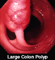
The lining of the colon and rectum is normally smooth. Sometimes, however, growths known as "colon polyps" appear on the lining of the large intestine. Varying in size from pinheads to growths several inches in diameter, polyps do not usually produce any symptoms. Contrary to popular belief, polyps affect both men and women equally. Most colon polyps are initially non-cancerous. Nevertheless, as colon polyps continue to grow, they often develop into cancer of the colon. Colon cancer has become quite common in our society. In fact, nearly 1 in every 20 adult Americans will develop colon cancer in their lifetime. Colon cancer is now the leading cause of cancer deaths in nonsmokers. Only lung cancer takes a greater toll. It is not widely known that colon cancer now kills more women than breast cancer and more men than prostate cancer. But, there is good news. Research has confirmed that the single best prevention for colon cancer is the early detection and removal of all colon polyps!!! In the past, colon polyps located beyond the range of the proctoscope could only be removed by a major abdominal operation. Of course, this procedure required about a week of hospitalization and over a month of time lost from work for recovery. Fortunately, medical technology now permits the simple removal of most colon polyps. Should a small polyp be discovered during your colonoscopy, the polyp can usually be removed immediately. This prevents a potential colon cancer while eliminating the need for major surgery.
..
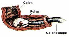
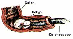
The doctor removes the polyp quite simply by placing a wire loop, or "snare," around the polyp's base and slowly tightening the loop. As the polyp is severed, a mild electric current cauterizes the tissue to prevent bleeding. There is no pain or any sensation as the polyp is removed. Polyp removal can usually be done on an outpatient basis. Moreover, since there is no incision, you may usually resume normal activity the next day.
..
After The Test
After an outpatient colonoscopy, you will be asked to rest awhile in the recovery room. Your companion will be asked to sit with you while the effects of the sedative begin to wear off. Once you are more alert, the doctor will meet with you to discuss the findings and any needed treatment. You will then be able to return home with your companion's assistance. However, since you will still be somewhat drowsy and uncoordinated, you will be taken directly to your car in a wheelchair by the office personnel. It is important that you go directly home; do not stop to eat along the way. Although you may resume a regular diet at home, you should eat lightly at first, and then gradually increase your intake of foods as tolerated. Since air was placed in the colon during the examination, you may experience the discomfort of mild "gas pains" for several hours until the gas is expelled. Because of the sedation's lingering effects, you should not drive, operate any machinery, drink alcohol, or engage in any vigorous activity for the remainder of the day. If the test is performed on an inpatient basis, you will be taken back to your hospital bed for recovery. If any specimens or polyps were extracted during the examination, the doctor will contact both you and your personal physician when the laboratory results become available. Further treatment or tests, if necessary, will be discussed at that time.
..
Is This Test Dangerous?
While every medical procedure involves some degree of risk, complications rarely occur in patients undergoing colonoscopy. With the advent of flexible fiberoptic instruments, this test has become a safe and simple method of directly examining the lower digestive tract. When performed by a physician who is specially trained and experienced in the procedure, the benefits of colonoscopy far exceed the risks.
..
Your doctor is a Gastroenterologist. In addition to standard medical training, he has received special instruction in diseases of the digestive system and has been thoroughly trained in the safe and proper operation of the colonoscope. The combination of his expertise and your cooperation should make this test as safe and simple as possible. However, as with all medical procedures, complications can occur.
..
The principal risks are perforation of the colon (a tear through the bowel wall) or bleeding. Although perforation generally requires surgery, certain cases may be treated with antibiotics and intravenous fluids. Bleeding may occur at the site of either a biopsy or polyp removal. Typically minor in degree, such bleeding may stop on its own or be controlled by cauterization. Occasionally, surgery is necessary. Fortunately, both perforation of the colon and bleeding are quite rare. Because bleeding may sometimes occur up to three weeks after a colon polyp has been removed, you should not plan to travel to any remote areas without medical access during this period of healing.
..
Other possible risks include drug reactions and complications related to other diseases that you may have. Consequently, you should inform your doctor of all allergic tendencies and medical problems. Occasionally, the site of the sedative injection may become inflamed and tender for a short time. This is not serious. If inflammation occurs, applying warm compresses to the area is usually helpful. While any of these complications may occur, it is well to remember that each of them occurs quite infrequently. Your doctor will be happy to discuss the above risks with you further, especially with regard to your particular situation and need for colonoscopy.
..
How About AIDS?
Due to the recent extensive coverage of AIDS by the media, some patients have expressed the concern that they might, in fact, contract AIDS through this examination. Be assured that this is not the case. All our instruments are thoroughly cleaned and undergo high-level disinfection after each use. Only techniques known to kill all disease-causing bacteria and viruses, including the hepatitis and AIDS virus, are employed in this process.
..
A Valuable Tool....
In summary, colonoscopy is a valuable tool for the diagnosis and treatment of many diseases of the large intestine. Even when x-rays are normal, the cause of symptoms such as rectal bleeding or change in bowel habits may be determined by colonoscopy. It is also useful in the diagnosis and follow-up care of patients with colitis. Through colonoscopy, the detection and removal of colon polyps, and thus the prevention of colon cancer, are made possible. Periodic colonoscopy is a valuable tool in monitoring patients with previous polyps, colon cancer, or a family history of colon cancer. In essence, colonoscopy is a safe and worthwhile procedure that is extremely well tolerated. If you have any questions about your need for colonoscopy, do not hesitate to ask your doctor.


To view information on another disease, click on Pancreatitis SOD Library.!

Pancreatitis SOD Library


..

Flexible Sigmoidoscopy

Flexible Sigmoidoscopy
..
Your doctor may have recommended that you undergo a special examination of your colon called Flexible Sigmoidoscopy, commonly called a "flex sig" Since your cooperation is essential for a successful and complete examination, it is important that you know exactly what is to be done and why. If, after reading this information, you have any additional questions concerning this procedure, ask your doctor.
..
What Is Flexible Sigmoidoscopy?
In the past, you may have experienced or heard of a proctoscope examination of the lower colon and rectum. This was performed with a short rigid hollow tube which allowed your doctor to examine only the last 10 inches of the large intestine. This device was often painful and, unfortunately, gave colon checkups a bad reputation as a very uncomfortable and embarrassing examination. Fortunately, recent progress in the field of fiberoptics has made colon exams much easier and more accurate. A new device, called a flexible sigmoidoscope, a 24 inch long thin flexible tube which contains a tiny color video camera is now used. This allows your doctor to perform a much more accurate and comfortable examination than the old "procto."
..
Why Do I Need This Test?
Flexible sigmoidoscopy can be done for a number of reasons, but the most common one is to help prevent colon cancer. It is now known that most colon cancers begin many years before as a small noncancerous growth called a polyp. But unfortunately, polyps cause no symptoms until they develop into colon cancer and by then it is too late. To minimize your personal risk of colon cancer, your colon must be periodically examined - just as one has periodic mammograms and prostate checkups. If a polyp is found during this exam and later removed, a potential colon cancer can be prevented. In other circumstances, flexible sigmoidoscopy can also be done to examine the left colon for signs of colitis, or to further evaluate symptoms such as diarrhea and rectal bleeding.
..
Is This Test Dangerous?
There is some risk to any medical procedure, but the frequency of serious complications during flexible sigmoidoscopy is quite low. When performed by an experienced physician, the benefits far exceed the risks. The combination of your doctor's experience and your cooperation should make this test as safe and simple as possible. However, as with all medical procedures, complications can occur. The principal risks are perforation of the colon (a tear through the bowel wall) or bleeding. A perforation usually requires surgery but may be managed with antibiotics and intravenous fluids in selected cases. Bleeding from flexible sigmoidoscopy is usually minor and stops on its own or can be controlled with treatment. Occasionally, blood transfusions and surgery are necessary. Fortunately, both of these problems are quite rare during flexible sigmoidoscopy - about 1 case in 8000 for perforation and 1 case in 17,000 for bleeding.
..
Scope Disinfection
With all the attention that AIDS and Hepatitis C have had in the media, some individuals have been concerned that they might contract a disease from these examinations. Be assured that this is not the case. All of the instruments are extensively cleaned and completely disinfected between each case with techniques that have been shown to effectively kill disease-causing bacteria and viruses including those which cause AIDS and hepatitis.
..
How Do I Prepare For The Exam?
Your cooperation is essential for a successful and complete examination. There are certain things you can do to help before the test begins. Your colon must be empty during flexible sigmoidoscopy so that your doctor's view is not blocked by particles of stool. This will require a special dietary and laxative preparation the day before the test. Detailed preparation instructions will be provided by the office staff. Be sure your doctor knows if you are taking insulin injections or Coumadin anticoagulants. Unless otherwise instructed, you may take all of your other routine medications as usual, even the day of the exam. Since anesthesia is not needed, a driver is not required.
..
Looking Inside
On the day of examination, wear comfortable easily-folded clothing that is simple to remove. Prior to the test, you will be asked to remove your clothing and to slip into a patient gown. Then, after a consent form is signed, you will be positioned comfortably on your left side on the padded examination table. The doctor will then do a rectal exam with a gloved, lubricated finger; then the lubricated scope will be gently inserted. This will not be painful, but is mildly uncomfortable for a few seconds. After the scope is positioned, the doctor will gently guide the flexible scope into the colon and examine the lining. To improve his view, your doctor will gently infuse air into the colon. This may cause a temporary sensation of abdominal fullness, cramping, or "gas," but the entire procedure should take less than three minutes. After The Test After the exam, you will be allowed to dress and have a glass of juice. The doctor will then explain to you exactly what was found and if any further tests or treatment will be necessary. Since this test does not require anesthesia, you will be able to immediately resume a regular diet and activities. Eat lightly at first, and then advance your diet as tolerated. Since air was placed in the colon during the examination, you may feel a little bloated and crampy until the extra air is expelled.
..
What If The Test Is Abnormal?
Don't panic. If a polyp is found, it is unlikely to be cancer - most are not. As a plus, most colon polyps can now be removed without the need for major abdominal surgery through a simple outpatient surgical procedure called colonoscopy. This more extensive "scope" examination is usually painless and does not require hospitalization. Most patients are back to work in a day or two. If more serious problems are found, the doctor will explain what medical or surgical forms of treatment are available.
..
What If The Test Is Normal?
Of course, since the flexible sigmoidoscope is only 2 feet long and the average colon is 5 feet long, not all of the colon is examined during this procedure. But, flexible sigmoidoscopy does allow examination of the area where the majority of colon cancer, polyps, and other problems are found. You must realize that this test is a simple screening exam and it is possible that problems further inside the colon may be missed. However, if you are in a low risk group, have no family history of colon cancer, no symptoms, nor signs of blood in the stool, the latest American Cancer Society guidelines merely recommend another flexible sigmoidoscopy every five years in the future and a stool test for hidden blood, or Hemoccult slide test, annually. However if you have symptoms or are in a higher risk group, your doctor may wish to alter these guidelines to your particular case. In Summary So as you have learned, flexible sigmoidoscopy is a safe, simple, and highly effective diagnostic technique that allows your doctor to directly inspect the lining of your left colon. As an individual, you can dramatically reduce your risk of getting colon cancer by having regular examinations before symptoms develop. If you are over 50 years old, consult with your family physician about periodic colon examinations. Prevention means taking charge of your health. Following these few simple steps can keep you healthy to enjoy the good life you have worked so hard to create. It's worth the effort....
..


To view information on another disease, click on Pancreatitis SOD Library.!

Pancreatitis SOD Library


..

MRI and MRCP

Magnetic Resonance Imaging (MRI)
...
What is an MRI?
Most radiology imaging studies use x-rays to visualize what lies inside the body. For example, a chest x-ray allows the doctor to see through the skin and study the heart and lungs. Modern x-rays are very safe, but do expose the body to some x-ray radiation. Magnetic resonance imaging (MRI) is a different method of looking inside the body. Instead of x-rays, the MRI scanner uses magnetism and radio waves to produce remarkably clear pictures. The powerful magnetic field causes the hydrogen ions in the body to become magnetized and line up in a certain order. The data received is analyzed and turned into an image by a high powered computer to create detailed image slices (cross sections) of your body. MRI can produce better soft-tissue images than standard x-rays and is better at distinguishing normal, healthy soft tissue from diseased tissue.
...
How does MRI differ from a CT scan?
One of the most basic differences between the two tests is that Computerized Tomography (CT) Scanning uses x-rays and MRI does not. A CT scan uses faster scanning times and can be performed in patients with pacemakers and other metallic implants. But a CT scan does expose the patient to x-rays and risks allergic reactions to intravenously administered iodine-containing dye. The MRI produces better images of the body's soft tissues and involves no x-rays or iodine dyes. But MRI scanning times are longer and difficult for patients who are not able to hold their breath. Patients with pacemakers and intra-cerebral aneurysm clips cannot be scanned by MRI.
...
Where Is an MRI performed?
MRI studies can be safely and accurately performed in a hospital radiology department, in a mobile MRI unit, or a freestanding MRI center.
..
Who performs the exam?
An MRI study is performed by a trained MRI technician under the supervision of a radiologist, a medical doctor trained in special imaging studies. The results of the test are analyzed by the radiologist and reported to your doctor within a few days.
..
Regular vs "Open" MRI
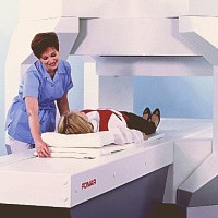
There are two types of MRI magnet machines: Closed and Open, based upon their shape. The standard "Closed" MRI is done in a narrow tube-like device about 2 feet in diameter and 6 feet to 8 feet long to optimize the images. Because of the small bore of the magnet, some patients experience claustrophobia and have difficulty in cooperating during the study. In the "Open" type, the large magnet that generates the image is generally suspended a couple of feet above the patient, and except for its supports, the unit is Open all around. If a patient is severely claustrophobic or over 300 pounds in weight, the doctor may suggest that the examination be done in an "Open" MRI unit because it has more room inside than a Closed magnet. Most Open units can accommodate patients up to 450 pounds in weight. The Open unit is more "patient friendly", but most radiologist feel that the Closed MRI operates much faster and produces a higher-resolution image with finer detail than the Open type. Closed magnets can be used for all MRI procedures, and Open magnets are used for more routine applications. Open MRI technology has improved over the years and may be adequate in many cases. Specialized studies such as MR angiography (MRA) and MR cholangiopancreatography (MRCP) can only be performed in Closed MRI machines.

Scheduling an MRI
An MRI is usually scheduled through the local hospital radiology department or a freestanding Open MRI center. In order to perform the study, they need an order for the study and an insurance referral from your physician. MRI is a non-invasive test and really without significant risks. One concern, however, is the "projectile effect", which involves the forceful attraction of metallic objects to the magnet. Because of this, there are several conditions under which MRI may not be safe. Patients should notify the receptionist or technologist prior to their appointment if they have any of the following:


  1. An implanted pacemaker, defibrillator ("AICD"), or heart valve
  2. An implanted pump device (such as an insulin or pain medication pump)
  3. An inner ear implant
  4. An aneurysm clip within the brain
  5. An intrauterine device (IUD)
  6. Metal in the eyes (at any time), or have ever been a metal worker of any kind
  7. Permanent tattoo eyeliner
  8. Currently pregnant
  9. Artificial joints or metallic plates
  10. Shrapnel Patients can safely undergo MRI with orthopedic hardware in their joints, such as a metallic plate or hip replacement. However, if the metal device is located close to the part of the body being examined, the images can be seriously degraded and useless.
..
MR cholangiopancreatography (MRCP)
This big word refers to a special test of the liver, bile ducts, and pancreas done using MRI. MRCP can produce images very similar as those obtained from the more invasive approach with ERCP (Endoscopic Retrograde Cholanigiopancreatography) without the added risk of pancreatitis, sedation, and perforation. However, image quality is less with MRCP and there is no way to correct whatever problem is found, as there is during ERCP. ERCP is best used when there is a high likelihood of gallstones obstructing the bile ducts or another blockage of the liver or pancreas. MRCP is of value in patients with a low probability of gallstones or obstruction in the bile ducts or pancreas, or in patients who are too sick for the anesthesia required for ERCP.
..
Preparing for an MRI
There is no special preparation for an MRI examination. There is no need for a change in daily routine. All prescription medications can be taken normally. However, patients undergoing MRI examination of the gallbladder and bile ducts (MRCP) will be asked to not eat for 12 hours prior to imaging. No special preparation is required for other body examinations. Patients are asked to bring the physician's order, insurance cards, referral forms, and any previous MRI, CT, or x-ray films relating to their exam. It is best to wear loose clothing without zippers or metallic parts. Elastic waistbands are suggested. During the exam The MRI technician explains the exam and answers any questions the patient may have. The patient may be asked to sign a consent form giving permission for the test and may be asked to change into a patient gown. Because MRI uses a powerful magnet, watches, metal objects in pockets, and credit cards with magnetic strips will not be permitted in the MRI room. Patients must also remove any other metallic objects such as jewelry, hairpins, eye glasses, wigs (if it has metallic clips), and non-permanent dentures. In a Closed MRI unit, the patient is positioned on a scanning table, head first, with arms at the side. The scanning table then slides into the magnet, covering the whole body. For clear pictures, the patient will be asked to hold very still, and in some cases, to hold their breath for up to 30 seconds. There is no pain or other sensation during the exam; however, an MRI is a noisy machine which produces intermittent humming, clicking and knocking sounds. Earplugs are available. Most MRI units also provide an assortment of music to help the patient relax. Patient are welcome to bring their own CD or cassette. There is a two-way intercom providing communication between patient and the technologist. For some studies such as MRCP, the radiologist will inject a "contrast agent" into a vein to improve the quality of the images. This material is injected into a vein in the arm. How long does an MRI exam take? The length of MRI examinations can vary from 15 minutes to 1 1/2 hours, averaging 45 minutes. Each test consists of several sequences or collections of data gathered over 2 to 10 minutes. After the exam There are no post-exam instructions. You may resume your normal diet and activities.
..
What about pregancy?
Although there are no known side effects of magnetism on the developing baby, it is recommended that a pregnant woman wait until the second trimester for MR imaging.

Getting the test results?
Because very large amounts of data are created during these studies, they can easily have hundreds of images that require hours of manipulation to interpret. The study will be read by a board-certified radiologist who sends the results to the referring physician who will notify the patient. Results are usually available within 72 hours.
..
What does an MRI cost?
The cost of an MRI study can range from $400 to more than $2,000, with a typical cost being about $800. Most health insurance, including Medicare, covers MRI testing. In summary MRI stands for Magnetic Resonance Imaging. An MRI offers a safe and efficient method of diagnosing many conditions, without the use of harmful x-rays. In many cases, MRI can lead to early detection and treatment of disease without surgery or biopsy. It is a non-invasive method of examining the soft tissue of the body including organs, muscles and tendons and requires little patient preparation. If you have any more questions about MRI, ask your doctor.
..
..

Magnetic Resonance Cholangiography (MRCP)
By: A. Alan White, M.D.
..
MRCP is a safe, non-invasive, easily performed and reliable procedure for evaluation of the bile ducts and the main pancreatic duct. No injections are required since the MR technique images the fluid normally present in the ducts. This is a procedure commonly utilized in major teaching institutions around the world.
..
Good quality examinations require a high field magnet, strong, fast gradients, special coils, and software that allows obtaining breath-hold heavily T-2 weighted images. All fluid containing structures will appear bright on these exams.
..
Techniques:
The patient fasts for 4 hours prior to the study to allow filling of the gallbladder and emptying of the stomach. The patient usually drinks about 100cc of a non absorbable iron containing liquid to decrease the signal of fluid in the stomach. The patient does not need IV contrast agents and there is no radiation exposure.
..
A series of images is then obtained during breath-holding periods of 2 to 25 seconds. Since the images are obtained rapidly, a series of images taken every few seconds through the distal duct can demonstrate contraction and relaxation of the distal common duct and sphincter of Oddi.
..
MRCP is based on a heavily T2 weighted pulse sequence which shows stationary fluids, such as bile, to tappear at high signal intensity whereas the surrounding liver and flowing blood generates little signal. As a result of this combination of imaging characteristics, MRCP provides optimal contrast between the hyperintense signal of the bile and the hypointense signal of background tissue.
..
Accuracy:
Detection of stones in the common duct by MRCP has an accuracy in the 90-95% range which is very comparable to ERCP. The accuracy of detection of the point of benign or malignant obstruction of the biliary duct or ducts is in the 90% range. Diagnosis of chronic pancreatitis involving the main duct is very comparable to ERCP with 90% correlation.
..
Indications for MRCP:

  • Patients where ERCP has failed, where ERCP carries a high risk and in patients where prior surgery makes access difficult are best studied by MRCP. In patients with low to moderate clinical suspicion of bile duct stones MRCP should be considered because of its high accuracy and lack of risk. With a high clinical suspicion of bile duct stones, ERCP is a logical choice since therapy can be instituted at the time of the procedure. Patients with acute pancreatitis have an increased risk of complication from ERCP and in patients with a low to moderate clinical suspicion of bile duct stones MRCP offers a no risk alternative.
  • Demonstration of ductal anatomy above a point of complete obstruction is another use of MRCP and is helpful in planning therapeutic intervention for bypass and drainage.
  • Suspected chronic pancreatitis, demonstration of anatomic variants such as pancreas divisum, aberrant cystic duct and other duct anomalies, sclerosing cholangitis, choledochal cysts, anastomic strictures and demonstration of post surgical anatomy are all uses for MRCP. Pre-operative evaluation of patients prior to laparoscopic cholecystectomy is currently under study. MRCP would be useful in those 10-15% of patients with common duct stones in helping plan the surgical procedure.
..
Advantages of MRCP Compared to ERCP:

  1. Non invasive (avoids complications of diagnostic ERCP or PTC)
  2. Usually no sedation required
  3. No iodinated intravenous contrast
  4. Rapid scan time
  5. No ionizing radiation
  6. Delineates ductal anatomy proximal to obstruction
  7. Delineates anatomy post biliary-enteric anastomosis
..
Contraindication:
The only contraindications are those of MRI in general including cardiac pacemakers, ferromagnetic aneurysm clips, intraorbital metallic foreign bodies and severe claustrophobia.
..
Summary:
MRCP has high diagnostic accuracy, is completely safe and avoids the risk of pancreatitis, bleeding and perforation associated with invasive techniques. The exam is well tolerated by most patients and diagnostic quality images can be obtained 99% of the time.
..

To view information on another disease, click on Pancreatitis SOD Library.!

Pancreatitis SOD Library


..

Secretin Testing

Secretin
From Wikipedia, the free encyclopedia

Secretin
Identifiers
Symbol
SCT
Entrez
6343
HUGO
10607
OMIM
182099
RefSeq
NM_021920
UniProt
P09683

Other data
Locus
Chr. 11 p15.5
Secretin is a peptide hormone produced in the S cells of the duodenum in the crypts of Lieberkühn.[1] Its primary effect is to regulate the pH of the duodenal contents via the control of gastric acid secretion and buffering with bicarbonate. It was the first hormone to be identified (see Discovery). In humans, the secretin peptide is encoded by the SCT gene.[2][3]

Discovery
In 1902, William Bayliss and Ernest Starling were studying how the nervous system controls the process of digestion.[4] It was known that the pancreas secreted digestive juices in response to the passage of food into the duodenum. They discovered (by cutting all the nerves to the pancreas in their experimental animals) that this process was not, in fact, governed by the nervous system. They determined that a substance secreted by the intestinal lining stimulates the pancreas after being transported via the bloodstream. They named this intestinal secretion secretin. Secretin was the first such "chemical messenger" identified. This type of substance is now called a hormone, a term coined by Bayliss in 1905.

Structure
Secretin is a linear peptide hormone, which is composed of 27 amino acids and has a molecular weight of 3055. A helix is formed in the amino acids between positions 5 and 13. The amino acids sequences of secretin have some similarities to that of glucagon, vasoactive intestinal peptide (VIP), and gastric inhibitory peptide (GIP). Fourteen of 27 amino acids of secretin reside in the same positions as in glucagon, 7 the same as in VIP, and 10 the same as in GIP.[5]
Secretin also has an amidated carboxyl-terminal amino acid which is valine.[6] The sequence of amino acids in secretin is: His-Ser-Asp-Gly-Thr-Phe-Thr-Ser-Glu-Leu-Ser-Arg-Leu-Arg-Asp-Ser-Ala-Arg-Leu-Gln-Arg-Leu-Leu-Gln-Gly-Leu-Val(NH2).[6]

Physiology


  • Production
    Secretin is synthesized in cytoplasmic secretory granules of S-cells which are found mainly in mucosa of duodenum, and smaller numbers in jejunum of small intestine.[7]
  • Stimulus
    Secretin is released into circulation and/or intestinal lumen in response to low duodenal pH that ranges between 4 and 4.5 depending on species.[8]
    It is the active form of prosecretin.This acidity is due to chyme, which contains hydrochloric acid, entering from the stomach via the pyloric sphincter.Secretin targets the pancreas, which cause the organ to secrete a bicarbonate-rich fluid that flows into the intestine. Bicarbonate ion is a base which neutralizes the acid, thus establishing a pH favorable to the action of other digestive enzymes to the small intestine and preventing acid burns[9] Other factors are also involved in the release of secretin such as bile salts and fatty acids which result in additional bicarbonate being added to the small intestine.[10] Secretin release is inhibited by H2 receptor antagonists which reduce gastric acid secretion. As a result, the pH in the duodenum increases above 4.5, and secretin cannot be released.[11]
  • Function
    Secretin stimulates the secretion of bile from the liver. It also increases watery bicarbonate solution from pancreatic duct epithelium. Pancreatic acinar cells have secretin receptors in their plasma membrane. As secretin binds to these receptors, it stimulates adenylate cyclase activity and converts ATP to cyclic AMP.[12] Cyclic AMP acts as second messenger in intracellular signal transduction and leads to increase in release of watery carbonate.It is known to promote the normal growth and maintenance of the pancreas.
    Secretin increases water and bicarbonate secretion from duodenal Brunner's glands in order to buffer the incoming protons of the acidic chyme.[13] It also enhances the effects of cholecystokinin to induce the secretion of digestive enzymes and bile from pancreas and gallbladder, respectively.
    It counteracts blood glucose concentration spikes by triggering increased insulin release from pancreas, following oral glucose intake.<[14]
    It also reduces acid secretion from the stomach by inhibiting gastrin release from G cells.[citation needed] This helps neutralize the pH of the digestive products entering the duodenum from the stomach, as digestive enzymes from the pancreas (eg, pancreatic amylase and pancreatic lipase) function optimally at neutral pH.[citation needed]
    In addition, secretin simulates pepsin secretion which can help break down proteins in food digestion. It also stimulates release of glucagon, pancreatic polypeptide and somatostatin.[8]
Uses
Secretin has been widely used in medical field especially in pancreatic functioning test. Secretin is either injected[15] or given through the tube that is inserted through nose, stomach then duodenum.[16] This test can provide information whether there are any abnormalities in pancreas which can be gastrinoma, pancreatitis or pancreatic cancer.
Extensive research has been conducted on the use of secretin to treat Autism. A "gut-brain" theory of autism proposes a link between the gastrointestinal disorders observed in many children with autism and their brain dysfunctions.[17]

References

  1. ^ Häcki WH (September 1980). "Secretin". Clin Gastroenterol 9 (3): 609–32. PMID 7000396.
  2. ^ Kopin AS, Wheeler MB, Leiter AB (March 1990). "Secretin: structure of the precursor and tissue distribution of the mRNA". Proc. Natl. Acad. Sci. U.S.A. 87 (6): 2299–303. PMID 2315322. PMC: 53674. http://www.pnas.org/cgi/pmidlookup?view=long&pmid=2315322.
  3. ^ Whitmore TE, Holloway JL, Lofton-Day CE, Maurer MF, Chen L, Quinton TJ, Vincent JB, Scherer SW, Lok S (2000). "Human secretin (SCT): gene structure, chromosome location, and distribution of mRNA". Cytogenet. Cell Genet. 90 (1-2): 47–52. PMID 11060443. http://content.karger.com/produktedb/produkte.asp?typ=fulltext&file=ccg90047.
  4. ^ Bayliss W, Starling EH (1902). "The mechanism of pancreatic secretion". J. Physiol. (London) 28: 325–353.
  5. ^ Williams, Robert L. (1981). Textbook of Endocrinology. Philadelphia: Saunders. pp. 697. ISBN 0-7216-9398-9.
  6. ^ a b DeGroot, Leslie Jacob (1989). J. E. McGuigan. ed. Endocrinology. Philadelphia: Saunders. pp. 2748. ISBN 0-7216-2888-5.
  7. ^ Polak JM, Coulling I, Bloom S, Pearse AG (1971). "Immunofluorescent localization of secretin and enteroglucagon in human intestinal mucosa". Scandinavian Journal of Gastroenterology 6 (8): 739–44. PMID 4945081.
  8. ^ a b Frohman, Lawrence A.; Felig, Philip (2001). "Gastrointestinal Hormones and Carcinoid Syndrome". in P. K. Ghosh and T. M. O’Dorisio. Endocrinology & metabolism. New York: McGraw-Hill, Medical Pub. Div. pp. 1326. ISBN 0-07-022001-8.
  9. ^ http://www.vivo.colostate.edu/hbooks/pathphys/endocrine/gi/secretin.html
  10. ^ Osnes M, Hanssen LE, Flaten O, Myren J (March 1978). "Exocrine pancreatic secretion and immunoreactive secretin (IRS) release after intraduodenal instillation of bile in man". Gut 19 (3): 180–4. PMID 631638. PMC: 1411891. http://gut.bmj.com/cgi/pmidlookup?view=long&pmid=631638.
  11. ^ Rominger JM, Chey WY, Chang TM (July 1981). "Plasma secretin concentrations and gastric pH in healthy subjects and patients with digestive diseases". Digestive diseases and sciences 26 (7): 591–7. PMID 7249893.
  12. ^ Gardner JD (1978). "Receptors and gastrointestinal hormones". in Sleisenger MH, Fordtran JS. Gastrointestinal Disease (2nd edition ed.). Philadelphia: WB Saunders Company.
  13. ^ Hall, John E.; Guyton, Arthur C. (2006). Textbook of medical physiology. St. Louis, Mo: Elsevier Saunders. pp. 800–801. ISBN 0-7216-0240-1.
  14. ^ Kraegen EW, Chisholm DJ, Young JD, Lazarus L (March 1970). "The gastrointestinal stimulus to insulin release. II. A dual action of secretin". J. Clin. Invest. 49 (3): 524–9. doi:10.1172/JCI106262. PMID 5415678.
  15. ^ "Human Secretin". Patient Information Sheets. United States Food and Drug Administration. 2004-07-13. http://www.fda.gov/cder/consumerinfo/druginfo/Human_Secretin.HTM. Retrieved on 2008-11-01.
  16. ^ "Secretin stimulation test". MedlinePlus Medical Encyclopedia. United States National Library of Medicine. http://www.nlm.nih.gov/medlineplus/ency/article/003892.htm#Definition. Retrieved on 2008-11-01.
  17. ^ "The Use of Secretin to Treat Autism". NIH News Alert. United States National Institutes of Health. 1998-10-16. http://www.nichd.nih.gov/news/releases/secretin.cfm. Retrieved on 2008-11-30.
See also
Secretin receptor

External links
Overview at colostate.edu
MeSH Secretin
Physiology at MCG 6/6ch2/s6ch2_17
[show]
v • d • eEndocrine system: hormones (Peptide hormones · Steroid hormones)
Endocrine glands
Hypothalamic-pituitary
Hypothalamus
GnRH · TRH · Dopamine · CRH · GHRH/Somatostatin
Posterior pituitary
Vasopressin · Oxytocin
Anterior pituitary
α (FSH, LH, TSH) · Prolactin · POMC (ACTH, MSH, Endorphins, Lipotropin) · GH
Adrenal axis
Adrenal cortex: aldosterone · cortisol · DHEAAdrenal medulla: epinephrine · norepinephrine
Thyroid axis
Thyroid: thyroid hormone (T3 and T4) · calcitoninParathyroid: PTH
Gonadal axis
Testis: testosterone · AMH · inhibin
Ovary: estradiol · progesterone · inhibin/activin · relaxin (pregnancy)Placenta: hCG · HPL · estrogen · progesterone
Other end. glands
Pancreas: glucagon · insulin · somatostatin
Pineal gland: melatoninThymus: Thymosin · Thymopoietin · Thymulin
Non-end. glands
digestive system: Stomach: gastrin · ghrelin · Duodenum: CCK · GIP · secretin · motilin · VIP · Ileum: enteroglucagon · Liver/other: Insulin-like growth factor (IGF-1, IGF-2)
Adipose tissue: leptin · adiponectin · resistin
Skeleton: Osteocalcin
Kidney: JGA (renin) · peritubular cells (EPO) · calcitriol · prostaglandinHeart: Natriuretic peptide (ANP, BNP)
Target-derived
NGF · BDNF · NT-3
[show]
v • d • eDigestive system, physiology: gastrointestinal physiology
Enteric nervous system
Meissner's plexus · Auerbach's plexus
Exocrine
Chief cells (Pepsinogen) · Parietal cells (Gastric acid, Intrinsic factor) · Goblet cells (Mucus)
Endocrine/paracrine
G cells (gastrin) · D cells (somatostatin) · ECL cells (Histamine)
enterogastrone: I cells (CCK) · K cells (GIP) · S cells (secretin)Enteroendocrine cells · Enterochromaffin cell · APUD cell
Border
Brunner's glands · Paneth cells · Enterocytes
Fluids
tract: Saliva · Gastric juice · Intestinal juiceaccessory: Bile · Pancreatic juice
Processes
upper GI: Swallowing · Vomiting
lower GI: Segmentation contractions · Migrating motor complex · Borborygmus · Defecation
either/both: Peristalsis (Interstitial cell of Cajal) · Gastrocolic reflexaccessory: Enterohepatic circulation
[show]
v • d • ePeptides: neuropeptides
Hypothalamic
Somatostatin - CRH - GnRH - GHRH - Orexins - TRH - POMC (ACTH, MSH, Lipotropin)
Gastrointestinal hormones
Cholecystokinin - Gastric inhibitory polypeptide - Gastrin - Motilin - Secretin - Vasoactive intestinal peptide
Other hormones
Vasopressin - Calcitonin -
Other
Angiotensin - Bombesin/Neuromedin B - Calcitonin gene-related peptide - Carnosine - Delta sleep-inducing peptide - FMRFamide - Galanin - Gastrin releasing peptide - Kinins (Bradykinin, Tachykinins ) - Neuromedin (B, N, U) - Neuropeptide Y - Neurophysins - Neurotensin - Opioid peptide - Pancreatic polypeptide - Pituitary adenylate cyclase activating peptide
[show]
v • d • eHormones: gastrointestinal hormones
CCK - EGF - GIP - Gastrin releasing peptide - Gastrins - Proglucagon - Motilin - Peptide YY -Prokineticin - Secretin - VIP

Retrieved from "http://en.wikipedia.org/wiki/Secretin"
Categories: Genes on chromosome 11 Peptide hormones Intestinal hormones Digestive system

To view information on another disease, click on Pancreatitis SOD Library.!
Pancreatitis SOD Library